
| Lot ID | Purity | Net Content | Endotoxin | Sterility |
|---|---|---|---|---|
| KLO0800314261 | 99.7 % | 86.4 mg (108.0%) | 33.3 EU/vial | Pass |
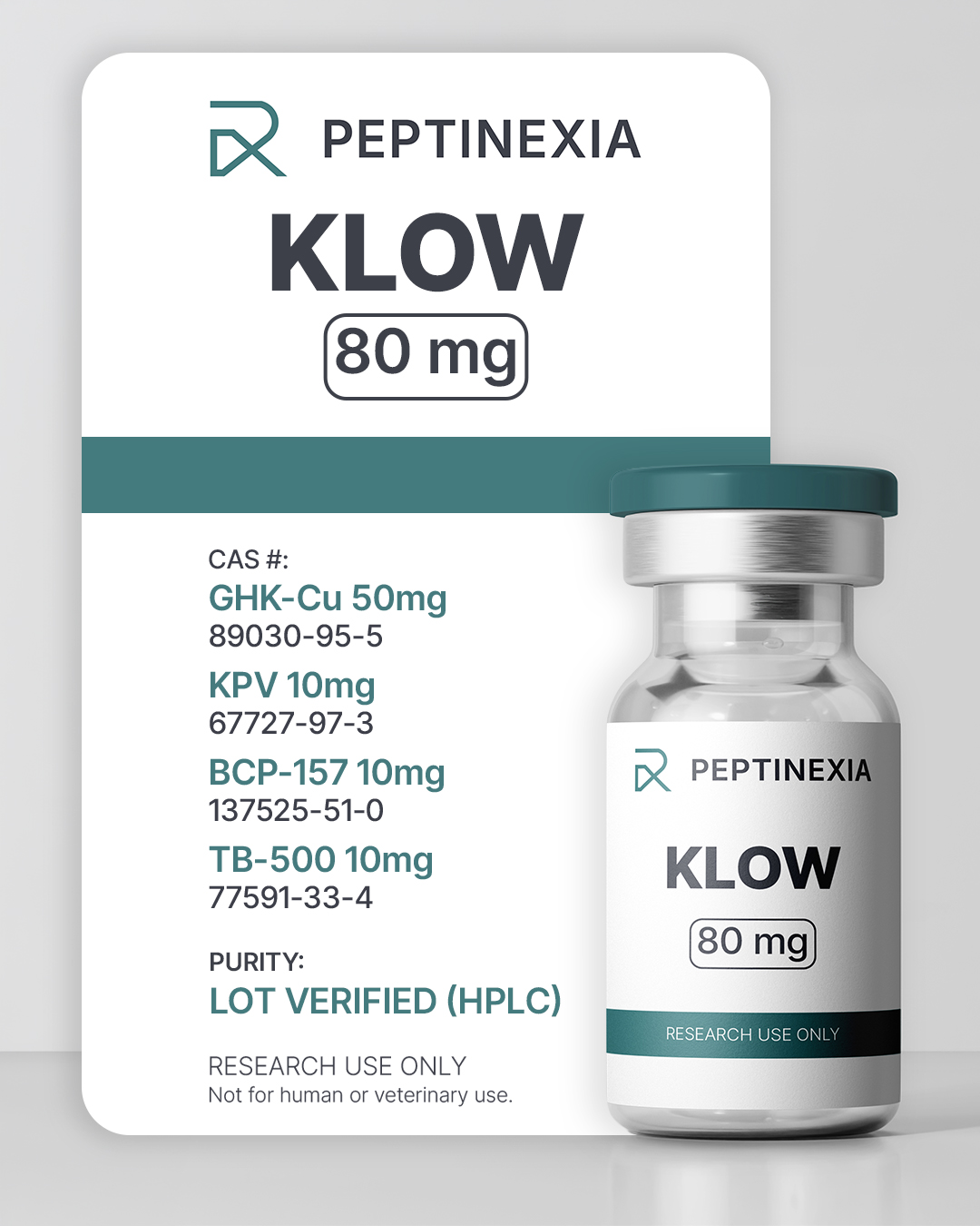
KLOW
KLOW is a multi-peptide research formulation combining GHK-Cu, BPC-157, TB-500 (thymosin beta-4), and KPV, investigated for coordinated cellular signaling, inflammatory modulation, and tissue remodeling mechanisms in preclinical research models.
$75.00
In stock
$75.00
1 - 2
vials
$71.25
(5% off)
3 - 5 vials
$67.50
(10% off)
6+ vials
What is KLOW?
KLOW is a multi-peptide research formulation combining GHK-Cu, BPC-157, thymosin beta-4 (TB-500), and KPV. Each component peptide has been independently studied for roles in cellular signaling, inflammatory modulation, and tissue remodeling processes. The formulation is designed for research investigating how coordinated peptide signaling may influence complex biological repair pathways.
Research Interest
KLOW is studied in experimental settings examining integrated cellular responses involving tissue remodeling, inflammatory signaling regulation, extracellular matrix dynamics, and vascular adaptation. Researchers explore how combining peptides with complementary biological activities may influence interconnected signaling systems compared with isolated peptide models.
Mechanisms Under Investigation
Research involving the individual components of KLOW suggests potential interactions across several biological pathways, including nitric oxide signaling, actin-mediated cellular migration, cytokine modulation, extracellular matrix regulation, and copper-dependent enzymatic activity. Investigations focus on how simultaneous pathway engagement may support coordinated cellular responses within complex tissue environments.
Current State of Research
While each peptide contained in KLOW has been studied independently in preclinical research, investigation of combined peptide formulations remains an emerging area of study. Ongoing research continues to explore how multi-peptide signaling approaches influence biological systems characterized by overlapping repair and regulatory pathways.
Lyophilized (Dry Powder) — Unopened Vials
Unopened lyophilized vials should be stored away from direct light and heat. For use within a few weeks, room temperature storage is acceptable. For storage over several months, refrigeration at 2–8°C (36–46°F) is recommended. For long-term storage, freezing best preserves peptide integrity.
When removing a vial from frozen storage, allow it to reach room temperature before opening to prevent condensation from introducing moisture into the vial.
Reconstitution
Reconstitute using bacteriostatic water (BAC). Inject the solution slowly down the inside wall of the vial rather than directly onto the peptide cake. Gently swirl until fully dissolved; do not shake. Vigorous shaking may cause foaming and mechanical stress to the peptide structure.
Reconstituted Vials
After reconstitution, store vials refrigerated at 2–8°C (36–46°F) and protected from light. Always use clean, sterile technique when accessing the vial to minimize contamination.
With proper refrigerated storage and aseptic handling, reconstituted peptide solutions commonly remain stable well beyond the frequently cited 28-day guideline, which pertains to the antimicrobial effectiveness of bacteriostatic water rather than the intrinsic peptide stability. Many researchers maintaining consistent sterile technique report usable stability in the 60–90 day range under controlled conditions.
General Guidelines
- Keep vials away from excessive heat and prolonged light exposure.
- Do not freeze after reconstitution.
- Discard any solution showing cloudiness, discoloration, or visible particulate matter.
- Label vials with the reconstitution date for tracking purposes.
Study 1: Copper Peptide GHK-Cu Modulates Gene Expression Associated with Tissue Remodeling
Authors: Pickart L., Margolina A.
Source: Journal of Aging Research
Scientific Findings
Genomic analysis demonstrated that GHK-Cu influences expression of genes involved in tissue remodeling, inflammatory signaling, antioxidant defense, and extracellular matrix regulation. The peptide exhibited broad regulatory effects across pathways associated with cellular repair and maintenance mechanisms.
Plain English Interpretation
Researchers found that GHK-Cu can influence groups of genes related to tissue structure and inflammation, suggesting a regulatory role in how cells coordinate repair-related processes.
Study 2: Pentadecapeptide BPC-157 Enhances Growth Hormone Receptor Expression in Tendon Fibroblasts
Authors: Chang CH et al.
Source: Molecules
Scientific Findings
This in-vitro study demonstrated increased growth hormone receptor expression and enhanced cellular proliferation in tendon fibroblasts exposed to BPC-157. Activation of downstream signaling pathways suggested increased responsiveness to endogenous repair signals.
Plain English Interpretation
Scientists observed that BPC-157 helped tendon cells respond more strongly to natural growth signals, supporting research into cellular repair mechanisms.
Study 3: Thymosin Beta-4 Promotes Cell Migration and Tissue Repair Responses
Authors: Malinda KM et al.
Source: Nature Medicine
Scientific Findings
Thymosin beta-4 stimulated endothelial and epithelial cell migration and accelerated wound closure in experimental models. Effects were linked to regulation of actin dynamics, a fundamental mechanism governing cellular movement and tissue remodeling.
Plain English Interpretation
Researchers found thymosin beta-4 helped cells move efficiently into damaged areas, an essential step in coordinated tissue repair responses studied in laboratory models.
Study 4: KPV Peptide Modulates Inflammatory Signaling Pathways
Authors: Catania A. et al.
Source: Peptides Journal
Scientific Findings
Research investigating the tripeptide KPV demonstrated modulation of inflammatory signaling pathways through interaction with melanocortin-related mechanisms. Experimental models showed reductions in pro-inflammatory cytokine activity and regulation of immune signaling responses.
Plain English Interpretation
Scientists studying KPV observed changes in inflammation-related signaling, suggesting the peptide may help regulate cellular responses involved in inflammatory processes.
Batch Details
ID
Content
Received Date
Lot Size
vials
Crimp Color
Cap Color
Testing Lab
Lab ID
Test Date
Peptide Purity
Net Peptide Content
Peptide-to-Excipients Ratio
Endotoxin
Sterility